 Home
Home
Character may be manifested in the great moments,
but it is made in the small ones.
Phillips Brooks
Home
Character may be manifested in the great moments,
but it is made in the small ones.
Phillips Brooks
You will use reverse transcriptase to prepare cDNA from total RNA. To confirm the quality of the isolated RNA, you will analyse RNA on a formaldehyde agarose gel.
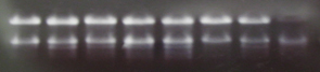
In both gel media, DNA is driven through the matrix by electric current. Smaller or more compact molecules pass through the matrix easier and migrate farther than large molecules. All linear DNA has the same charge per unit length and linear pieces migrate according to size. Plasmid DNA preparations contain three types of DNA conformations: linear, relaxed circular (or nicked) and supercoiled. Only the linear form can be used to estimate the size of the molecule. Usually, but not always, the supercoiled runs fastest, linear next, then the relaxed circular. A carefully prepared sample will be mostly supercoiled. The range of sizes separated in a gel is controlled by the % of agarose or %T of acrylamide in the gel.
Resolution Versus Matrix Concentration
Agarose |
Useful for Range of Linear dsDNA Molecules (kb) |
Acrylamide |
Useful for Range of Linear dsDNA Molecules (bp)* |
|
0.3 0.6 0.7 0.9 1.2 1.5 2.0 |
5 - 60 1 - 20 0.8 - 10 0.5 - 7 0.4 - 6 0.2 - 3 0.1 - 2 |
3.5 5.0 8.0 12.0 15.0 20.0 |
100 - 1000 75 - 500 50 - 400 35 - 250 20 - 150 5 - 100 |
The mobility is proportional to the voltage applied at low voltage but increasing voltage decreases the resolution of larger fragments of DNA. A general guideline for agarose gels in 1xTBE is 5V/cm maximum for resolving fragment lengths greater than 2 kb. The distance between the electrodes serves as the length in the calculation. Higher voltages increase the temperature of the gel causing increased band width and distortion of the lanes. The agarose can also melt, especially the low melting point agarose sometimes used when DNA is to be recovered from the gel. Voltages for acrylamide gels are generally twice the recommended voltage of the agarose gels but it is important to check manufacturer recommendations for the gel or for the electrophoretic equipment.
The mobility is also influenced by the choice of buffer systems. Besides the Tris Borate EDTA, pH 8.3 (TBE) buffer used in our experiments, a Tris Acetate EDTA buffer (TAE) is preferred by some. The TAE buffer shifts the range of resolution toward higher fragment lengths.
Denaturing gels can also be run to separate fragments as single stranded DNA. Gels can be run at high pH (30 mM NaOH) or in neutral buffers when glyoxal is added to the gel and the running buffer. Only the glyoxal system is suitable for RNA separations. Why is there a restriction of systems for RNA separations?
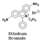
The nucleic acids are visualized with ethidium bromide (EtBr). This fluorescent dye, which contains a tricyclic planar group, intercalates between stacked base pairs of nucleotides and, in this environment, fluoresces when excited with ultraviolet light; the fixed position of the planar group and its close proximity to the bases causes dye bound to DNA to display increased fluorescent yield compared to free dye.
The tracking dye combined with the DNA samples contains bromophenol blue and xylene cyanol for use in visually monitoring electrophoresis and glycerol to make the sample dense enough sink to the bottom of the well. The stock solution is designed to be diluted about six fold in the sample. Bromophenol blue runs about the same size as a linear double-stranded DNA molecule of 300 base pairs in length in 1X TBE on a gel of 1% agarose. In low percentage gels of 0.4% agarose, the dye can emulate a 1000bp fragment. Remember not to run this dye off the bottom of the gel when you are trying to analyze small fragments. Xylene cyanol runs about the same as a linear double-stranded DNA molecule of 4kb in a 1% agarose gel.
Copyright, Acknowledgements,
and Intended Use
Created by B. Beason (bbeason@rice.edu), Rice University, 23 July 2003
Updated 19 March 2015