 Home
Home
Plasmids are small circles (usually less than 15 kb) of double stranded DNA maintained in some bacteria because they confer an advantage to the cells such as resistance to an antibiotic. The ease with which this DNA can be isolated and manipulated accounts for the widespread use of plasmids in molecular biology for a variety of tasks. Although plasmids occur naturally, the ones used in research have been engineered to prevent natural transfer between bacteria to reduce the spread of antibiotic resistance.
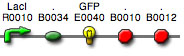
Some portions of the plasmid are essential for replication and maintenance. The ORI site directs the replication of the plasmid in bacteria and is essential on any plasmid. The left and right borders (LB and RB) are important for expressing . A special gene confers antibiotic resistance and permits selection of positive bacterial transformants.
The two most common methods for lysing bacteria are by alkali (1) and boiling (2,3). Commercial preparation kits are available for both small (miniprep) and large (maxiprep) scale DNA isolations. The miniprep is expected to yield 10-20 µg of plasmid DNA from 1-3 ml of culture. A maxiprep may provide up to a milligram of plasmid DNA from 100-300 ml of culture. These kits employ a variety of methods ranging from special solvents to customized columns with a wide range of cost.
The method used in this course employs initial alkaline lysis and produces DNA suitable for digestion with most restriction enzymes. The bacteria are harvested and rinsed to remove the media prior to being lysed with NaOH/SDS. The alkaline SDS solution breaks open the bacterial cells and prevents the activity of endogenous DNase activity. The NaOH denatures many of the cell components, including the DNA, so the pH must be quickly lowered in the next step. The chromosomal DNA, being very large and already somewhat fragmented, does not have the time to renature properly but forms a gel-like structure through small stretches of annealing. The chromosomal DNA (and cell membrane fragments) are then removed by centrifugation. The plasmid DNA remains soluble due to quick reannealing because the small circles of DNA are intertwined. During the initial stage of the preparation, special care should be taken to mix the solutions gently by inversion only. Avoid vigorous shaking or vortexing as this can shear the chromosomal DNA leading to contamination of the plasmid preparation. The supernatant is neutralized and brought to a high salt concentration with ammonium or potassium acetate and plasmid DNA is recovered by precipitation or by adsorption.
The plasmid DNA is bound to a silica membrane in high salt and then is eluted from the matrix in water or Tris-EDTA (TE) buffer. This rapid preparation should be essentially free of RNA contamination. The presence of RNA can interfere with digestions, electrophoresis gel analysis and A260 quantitation.
![]() The bacteria used
in this experiment are resistant to the antibiotic kanamycin
and should not be allowed to escape into the "wild." Prudent
lab technique must always be observed while manipulating these
research tools.
The bacteria used
in this experiment are resistant to the antibiotic kanamycin
and should not be allowed to escape into the "wild." Prudent
lab technique must always be observed while manipulating these
research tools.
![]() AFTER the
cells are lysed, such as by the addition of NaOH/SDS in
the plasmid prep, this and subsequent solutions are not
considered biohazardous and do not need to be decontaminated.
Tips, tubes, and other materials used after the lysis step
can be placed in regular trash.
AFTER the
cells are lysed, such as by the addition of NaOH/SDS in
the plasmid prep, this and subsequent solutions are not
considered biohazardous and do not need to be decontaminated.
Tips, tubes, and other materials used after the lysis step
can be placed in regular trash.
Copyright, Acknowledgements,
and Intended Use
Created by B. Beason (bbeason@rice.edu), Rice University, 6 February 2008
Updated 22 April 2009